Topographic sketch. Coronal view of bilateral DRT (orange). Patient
Por um escritor misterioso
Last updated 11 abril 2025


Journal of Comparative Neurology, Systems Neuroscience Journal

Subthalamotomy for Parkinson's disease: clinical outcome and topography of lesions

Abstracts for the UIP XIX World Congress of Phlebology, 12–16 September 2022, Istanbul, Turkey, 2022
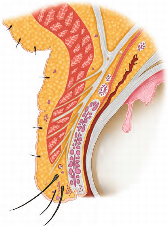
Anatomy of the Ocular Adnexa, Orbit, and Related Facial Structures
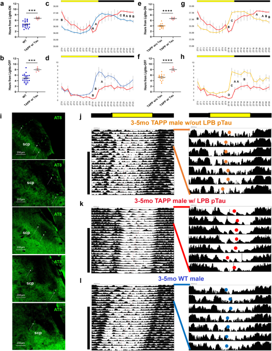
A brainstem to circadian system circuit links Tau pathology to sundowning-related disturbances in an Alzheimer's disease mouse model

ebook dentistry by Sach b m - Issuu

ECVP 2002 Abstract Supplement (complete) - Perception

Crossing nerve transfer drives sensory input–dependent plasticity for motor recovery after brain injury

PDF) Three-dimensional LASIK flap thickness variability: Topographic central, paracentral and peripheral assessment, in flaps created by a mechanical microkeratome (M2) and two different femtosecond lasers (FS60 and FS200)
Recomendado para você
-
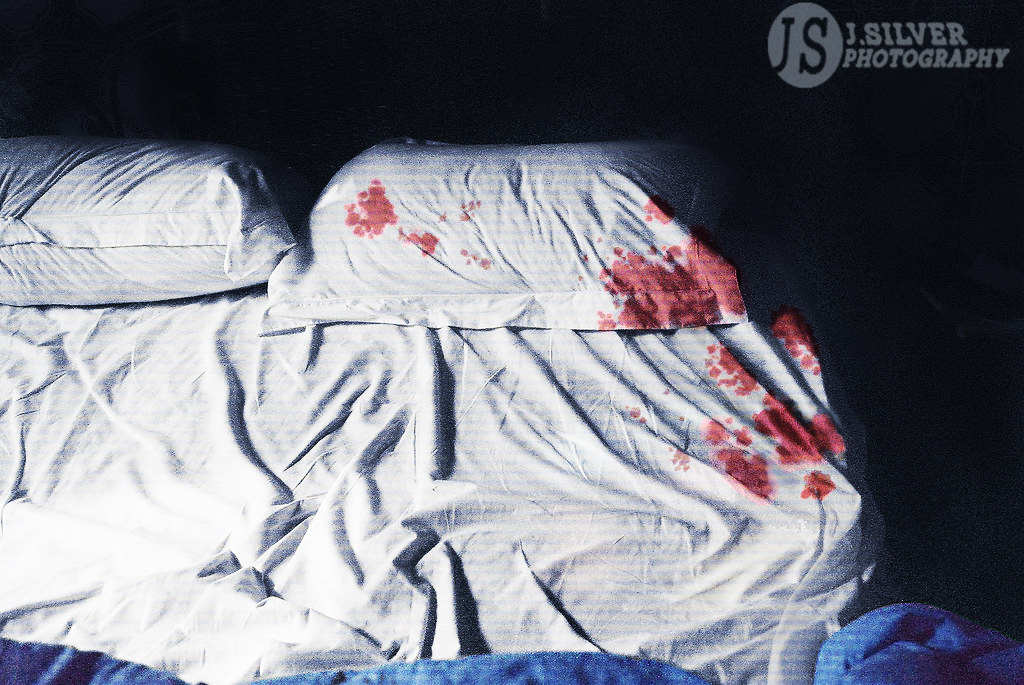
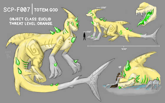
 SCP-007-PT - SCP International11 abril 2025
 -
 International Collaborations Hub - SCP International11 abril 2025
 -
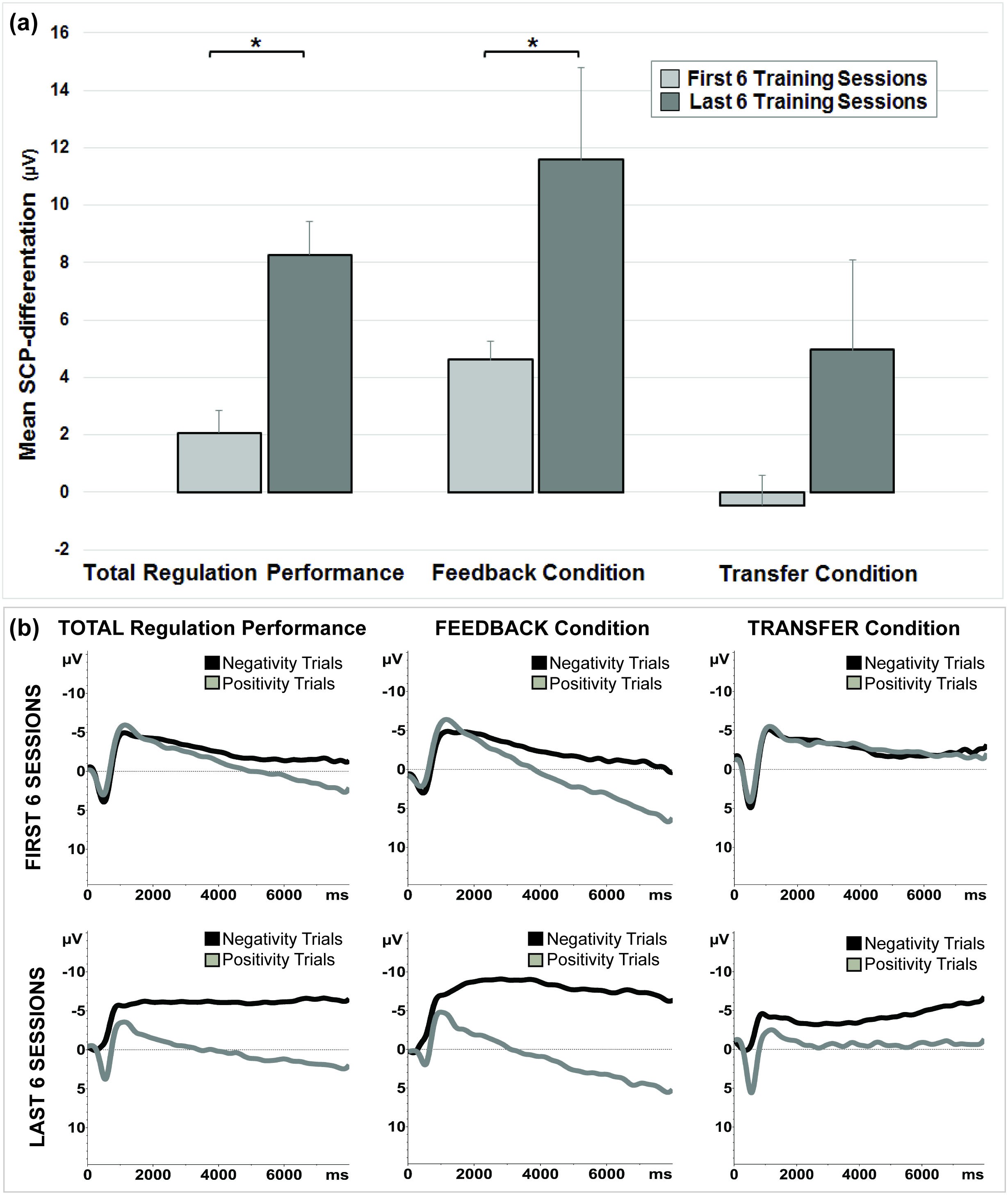 Brain self-regulation in criminal psychopaths11 abril 2025
 -
 Minecraft SCP foundation: building SCP-007 pt 111 abril 2025
 -
 SCP's I made Minecraft Collection11 abril 2025
 -
 SCP-006-PT - Fundação SCP11 abril 2025
 -
 More than just the spice of life: Using variety as a signal for change and diversification - Kahn - 2022 - Consumer Psychology Review - Wiley Online Library11 abril 2025
 -
![SCP-005 – SCP [ARCHIVOS FILTRADOS] – Podcast – Podtail](https://is1-ssl.mzstatic.com/image/thumb/Podcasts124/v4/ff/63/de/ff63de8f-c222-6fc5-7058-728b65394237/mza_5135098148032367375.jpg/250x250bb.jpg) SCP-005 – SCP [ARCHIVOS FILTRADOS] – Podcast – Podtail11 abril 2025
SCP-005 – SCP [ARCHIVOS FILTRADOS] – Podcast – Podtail11 abril 2025 -
 Used Eaton (Moeller) xPole Circuit Breaker System for Lab Distillations for Sale at Chemistry RG11 abril 2025
 -
 Explore the Best Foolishgamers Art11 abril 2025
você pode gostar
-
 The SCP Experience on Apple Podcasts11 abril 2025
 -
Kawaki Legacy - Imagine timeskip Boruto with susanoo and the11 abril 2025
-
 Freddie Prinze Jr Shares the Story Behind Kanan's Cameo in Star Wars: The Rise of Skywalker - IGN11 abril 2025
 -
 Cyberpunk: Edgerunners Shows Off David, Rebecca and Lucy Figures, 2024 Release Date - Anime Corner11 abril 2025
 -
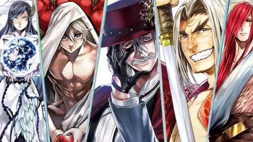 Create a Melhores Personagens de Shuumatsu no Valkyrie Tier List - TierMaker11 abril 2025
 -
 Pokemon Sword And Shield Ultimate All Pokemon Cheats Legendary Ultra Beasts And Many More11 abril 2025
 -
 minx on X: fun announcement coming soon 😌11 abril 2025
 -
 Download free Tiny Eddsworld Tord Minimal Background Wallpaper11 abril 2025
 -
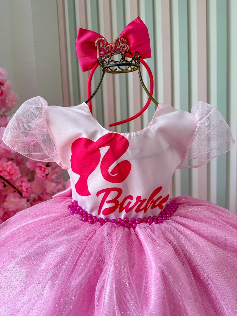 Vestido Barbie Branco e Rosa Tule Luxo Menina Infantil11 abril 2025
 -
 revengers Minecraft Skins11 abril 2025
