Cells, Free Full-Text
Por um escritor misterioso
Last updated 01 abril 2025
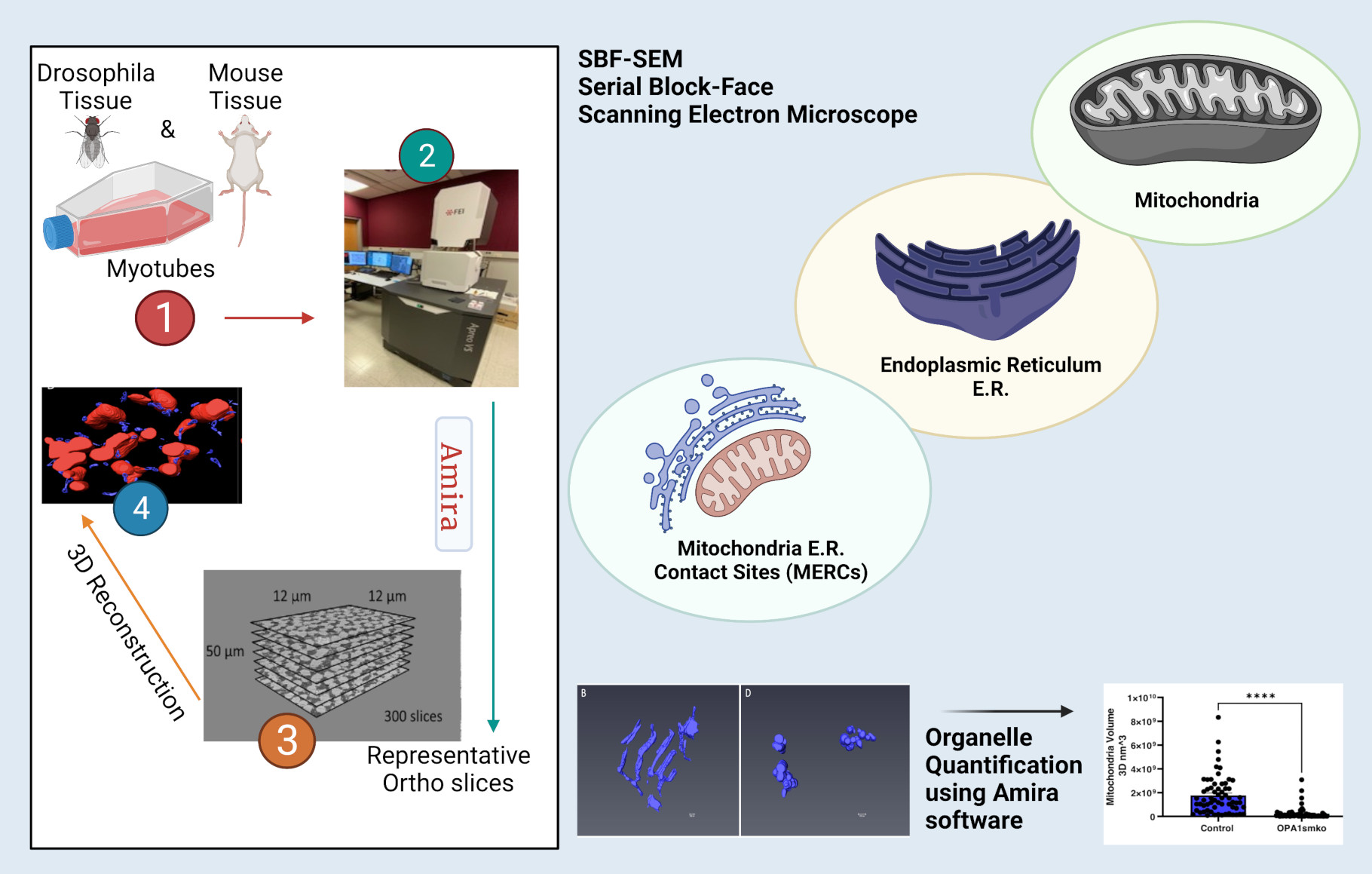
High-resolution 3D images of organelles are of paramount importance in cellular biology. Although light microscopy and transmission electron microscopy (TEM) have provided the standard for imaging cellular structures, they cannot provide 3D images. However, recent technological advances such as serial block-face scanning electron microscopy (SBF-SEM) and focused ion beam scanning electron microscopy (FIB-SEM) provide the tools to create 3D images for the ultrastructural analysis of organelles. Here, we describe a standardized protocol using the visualization software, Amira, to quantify organelle morphologies in 3D, thereby providing accurate and reproducible measurements of these cellular substructures. We demonstrate applications of SBF-SEM and Amira to quantify mitochondria and endoplasmic reticulum (ER) structures.

Cells, Free Full-Text
Antibodies, Free Full-Text
HIV-1 cell-to-cell transmission and broadly neutralizing antibodies, Retrovirology
Cells, Free Full-Text

Sequencing of Circulating Cell-free DNA during Pregnancy
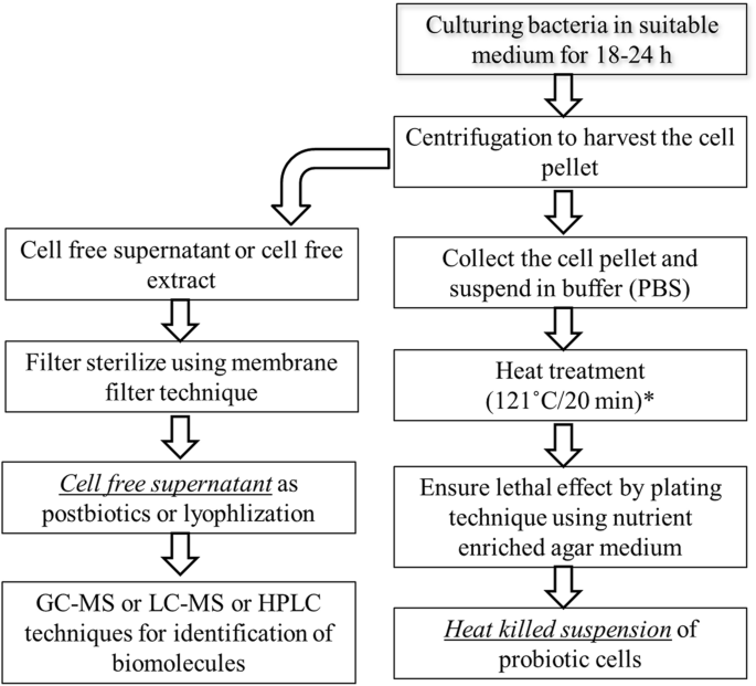
Postbiotics-parabiotics: the new horizons in microbial biotherapy and functional foods, Microbial Cell Factories

Cell-free Macromolecular Synthesis
Ds Kumar Strömungsmechanik Pdf Kostenloser Download - Colaboratory

PDF] Mesenchymal Stem Cell-Derived Exosomes as New Remedy for the Treatment of Neurocognitive Disorders

Five-Year Outcomes for Refractory B-Cell Lymphomas with CAR T-Cell Therapy
Recomendado para você
-
 Clicker Games - Play Free Clicking Games Online01 abril 2025
 -
 Mouse Accuracy - Mouse Accuracy and Pointer Click Training01 abril 2025
 -
 Mouse Accuracy & Reaction Timing practice01 abril 2025
 -
 How to Improve 3D-Printed Shape Accuracy? -Sharp Corner (XY Axis) – Raise3D: Reliable, Industrial Grade 3D Printer01 abril 2025
 -
 Free 3D file TEST SHAPE Finalmouse Ultralight Medium ZS-F2 Wireless 3D Printed Mouse 🐁・Model to download and 3D print・Cults01 abril 2025
 -
 New 3-D printer is 10 times faster than commercial counterparts, MIT News01 abril 2025
 -
 3Dconnexion SpaceMouse Pro 3D Mouse (3DX-700040) : Electronics01 abril 2025
 -
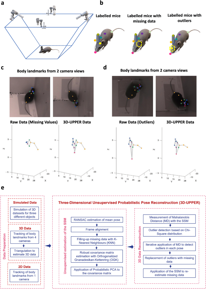 Three-dimensional unsupervised probabilistic pose reconstruction (3D-UPPER) for freely moving animals01 abril 2025
 -
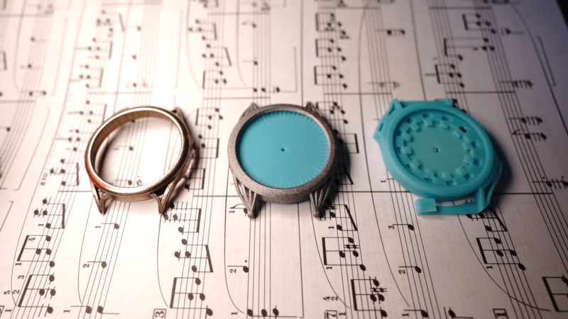 Put 3D Metal Printing Services To The Test, By Making A Watch01 abril 2025
 -
 Biomedicines, Free Full-Text01 abril 2025
você pode gostar
-
Watch Puppet Master Streaming Online01 abril 2025
-
 Lego Sonic The Hedgehog gets four amazing new sets and minifigures01 abril 2025
 -
 15 Words That Aren't As Straightforward As They Look01 abril 2025
 -
 The Revisited Meninas - The Incredible Tributes to Velasquez01 abril 2025
 -
 Yahari ore no seishun rabukome wa machigatteiru. (2013) Japanese movie poster01 abril 2025
 -
![Comentário ENEM 2010]](https://comentario.fariasbrito.com.br/upload/1292451182.204075391.gif) Comentário ENEM 2010]01 abril 2025
Comentário ENEM 2010]01 abril 2025 -
 Miguel Cariad, Ordem Paranormal Wiki01 abril 2025
 -
 locket heart Animated Gif Maker - Piñata Farms - The best meme generator and meme maker for video & image memes01 abril 2025
 -
 Todos Codigos Gta Sandres Playstation Ps301 abril 2025
 -
 SoulSeek Qt 2017.2 - Download for PC Free01 abril 2025