Figure 1. [The normal human retina fundus]. - Webvision - NCBI
Por um escritor misterioso
Last updated 10 abril 2025
![Figure 1. [The normal human retina fundus]. - Webvision - NCBI](https://www.ncbi.nlm.nih.gov/books/NBK554706/bin/Archetecture_Fovea-Image006.jpg)
The normal human retina fundus photo shows the optic nerve (right), blood vessels and the position of the fovea (center).
![Figure 1. [The normal human retina fundus]. - Webvision - NCBI](https://www.ncbi.nlm.nih.gov/books/NBK11556/bin/factsf2a.gif)
Facts and Figures Concerning the Human Retina - Webvision - NCBI Bookshelf
![Figure 1. [The normal human retina fundus]. - Webvision - NCBI](https://pub.mdpi-res.com/symmetry/symmetry-15-01631/article_deploy/html/images/symmetry-15-01631-g002.png?1692867486)
Symmetry, Free Full-Text
![Figure 1. [The normal human retina fundus]. - Webvision - NCBI](https://journals.sagepub.com/cms/10.1177/15353702211022674/asset/images/large/10.1177_15353702211022674-fig1.jpeg)
Interpretation of anatomic correlates of outer retinal bands in optical coherence tomography - Xincheng Yao, Taeyoon Son, Tae-Hoon Kim, David Le, 2021
![Figure 1. [The normal human retina fundus]. - Webvision - NCBI](https://www.cell.com/cms/asset/caee552e-ec98-489a-bfee-13674a9775ca/fx1.jpg)
Cell therapy with hiPSC-derived RPE cells and RPCs prevents visual function loss in a rat model of retinal degeneration: Molecular Therapy - Methods & Clinical Development
![Figure 1. [The normal human retina fundus]. - Webvision - NCBI](https://www.ncbi.nlm.nih.gov/books/NBK11556/bin/factsf5.gif)
Facts and Figures Concerning the Human Retina - Webvision - NCBI Bookshelf
![Figure 1. [The normal human retina fundus]. - Webvision - NCBI](https://www.ncbi.nlm.nih.gov/books/NBK11533/bin/sretinaf16.gif)
Simple Anatomy of the Retina - Webvision - NCBI Bookshelf
![Figure 1. [The normal human retina fundus]. - Webvision - NCBI](http://webvision.med.utah.edu/wp-content/uploads/2011/01/OCTmacula.jpg)
Simple Anatomy of the Retina by Helga Kolb – Webvision
![Figure 1. [The normal human retina fundus]. - Webvision - NCBI](https://pub.mdpi-res.com/symmetry/symmetry-15-01631/article_deploy/html/images/symmetry-15-01631-g007.png?1692867492)
Symmetry, Free Full-Text
![Figure 1. [The normal human retina fundus]. - Webvision - NCBI](https://media.springernature.com/m685/springer-static/image/art%3A10.1038%2Fs41598-021-04323-3/MediaObjects/41598_2021_4323_Fig3_HTML.jpg)
Asymmetry between right and left fundus images identified using convolutional neural networks
![Figure 1. [The normal human retina fundus]. - Webvision - NCBI](https://onlinelibrary.wiley.com/cms/asset/7f31a594-e67e-4087-8b31-ab0dd53da982/aos14249-fig-0015-m.jpg)
Subretinal surgery: functional and histological consequences of entry into the subretinal space - Sørensen - 2019 - Acta Ophthalmologica - Wiley Online Library
![Figure 1. [The normal human retina fundus]. - Webvision - NCBI](https://journals.sagepub.com/cms/10.1177/1535370218816517/asset/images/large/10.1177_1535370218816517-fig5.jpeg)
Functional optical coherence tomography of retinal photoreceptors - Xincheng Yao, Taeyoon Son, Tae-Hoon Kim, Yiming Lu, 2018
![Figure 1. [The normal human retina fundus]. - Webvision - NCBI](https://media.springernature.com/full/springer-static/image/art%3A10.1038%2Fs41467-019-12917-9/MediaObjects/41467_2019_12917_Fig1_HTML.png)
Single-nuclei RNA-seq on human retinal tissue provides improved transcriptome profiling
![Figure 1. [The normal human retina fundus]. - Webvision - NCBI](https://eophtha.com/images/uploads/15974739483541657755f37849ce2a63.jpg)
Anatomy of Retina
![Figure 1. [The normal human retina fundus]. - Webvision - NCBI](https://www.frontiersin.org/files/Articles/1106728/fpubh-11-1106728-HTML/image_m/fpubh-11-1106728-g001.jpg)
Frontiers Biomechanical homeostasis in ocular diseases: A mini-review
![Figure 1. [The normal human retina fundus]. - Webvision - NCBI](https://www.ncbi.nlm.nih.gov/books/NBK554060/bin/466648_1_En_8_Fig1_HTML.jpg)
Fig. 8.1, [Cross-sectional OCT image of human retina with the corresponding cellular structures]. - High Resolution Imaging in Microscopy and Ophthalmology - NCBI Bookshelf
Recomendado para você
-
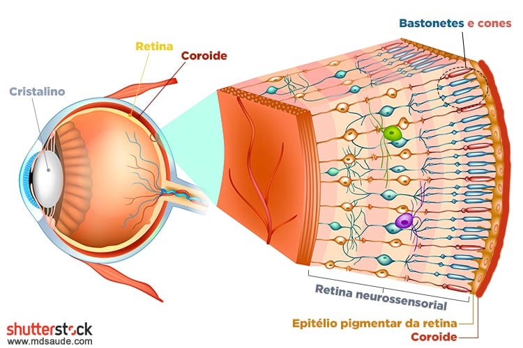
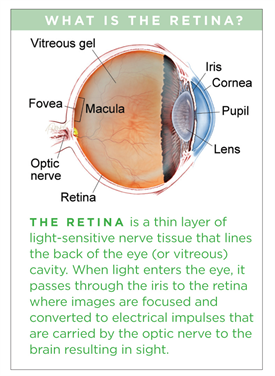
:max_bytes(150000):strip_icc()/GettyImages-308783-003-56acdcd85f9b58b7d00ac8e8.jpg) Retina: Anatomy, Function, and Treatment10 abril 2025
Retina: Anatomy, Function, and Treatment10 abril 2025 -
 Retinal Diseases: Types, Causes, Symptoms, Treatment, Outlook10 abril 2025
 -
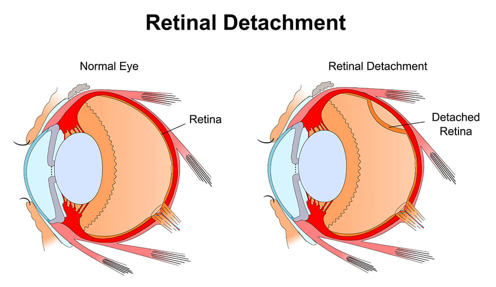 Retinal Detachment - Vitreo-Retinal Consultants10 abril 2025
 -
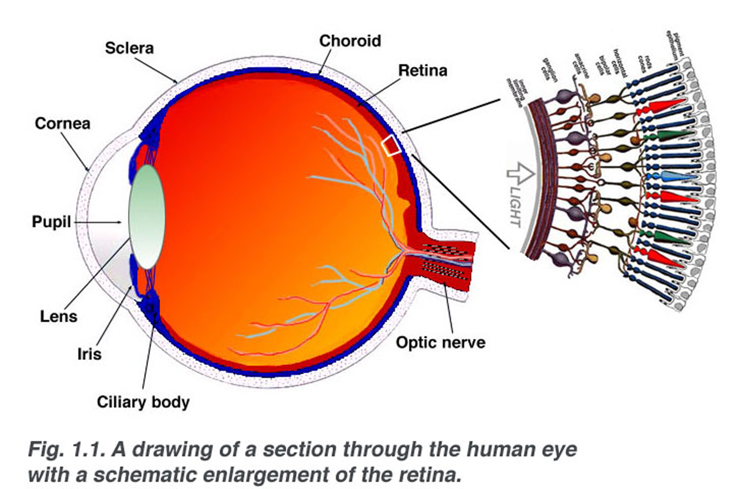 Simple Anatomy of the Retina by Helga Kolb – Webvision10 abril 2025
 -
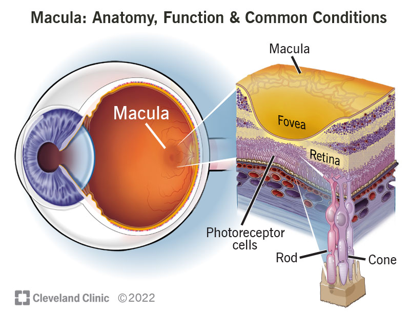
Macula: Anatomy, Function & Common Conditions10 abril 2025
-
 Descolamento de retina: sintomas e tratamento10 abril 2025
 -
 Retinal diseases - Symptoms and causes - Mayo Clinic10 abril 2025
 -
 Macular Edema - Patients - The American Society of Retina Specialists10 abril 2025
 -
 Retinal Tear vs. Retinal Detachment - Monterey, CA - Salinas10 abril 2025
 -
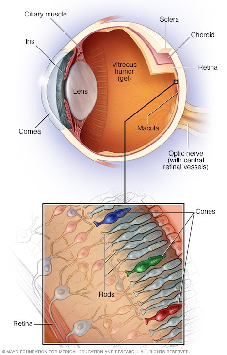
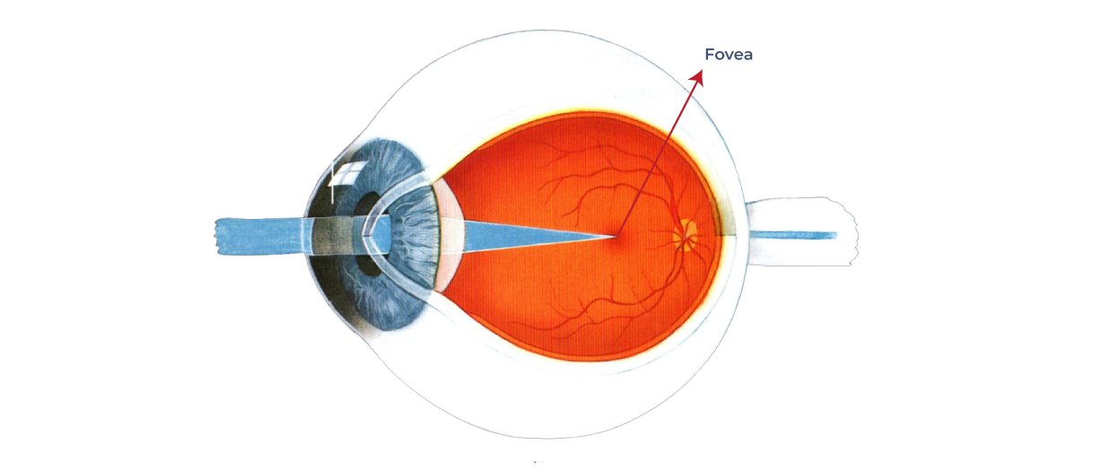 What is the fovea? – Front Range Retina10 abril 2025
você pode gostar
-
Ben 10: Ultimate Alien Characters List w/ Photos10 abril 2025
-
Aiya on X: Majutsushi Orphen Hagure Tabi Ep 1 I hope Orphen can10 abril 2025
-
 When Is 'Fire Force' Season 3 Coming Out?10 abril 2025
 -
 CapCut Pro APK Mod 10.3.0 Download grátis (Premium desbloqueado)10 abril 2025
 -
 Vanitas Anime Pop Art Greeting Card for Sale by Mitsugoshi10 abril 2025
 -
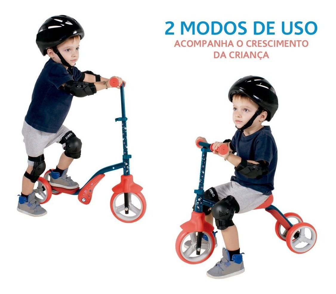 Patinete Infantil 2 Em 1 Vira Triciclo Bibiciclo Bel10 abril 2025
 -
 Check out the most anticipated games of 202210 abril 2025
 -
 Animação Sem Direitos Autorais10 abril 2025
 -
 How to do The best Six Sigma Green belt project Kick off meeting – Six Sigma Mania10 abril 2025
 -
 Little Buddy 1402 Kirby Adventure All Star Collection10 abril 2025